Malton Equine Veterinary Services
Mare Reproductive Services
In equine veterinary medicine, the ability to determine foetal gender is a valuable aspect of reproductive care, aiding breeders in their strategic breeding programs. Through the use of advanced imaging techniques, we offer precise foetal gender determination at different stages of gestation
Early Gender Determination via Transrectal Ultrasonography:
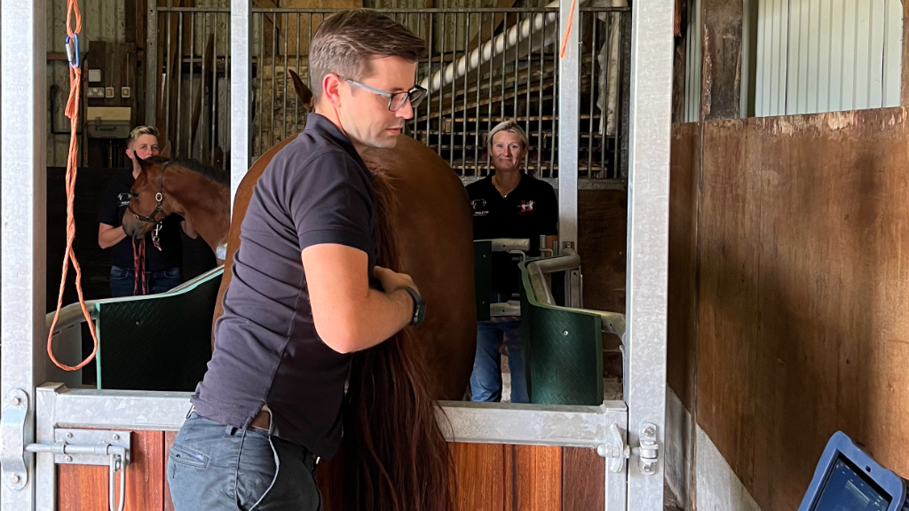
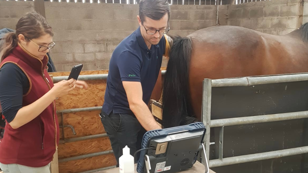
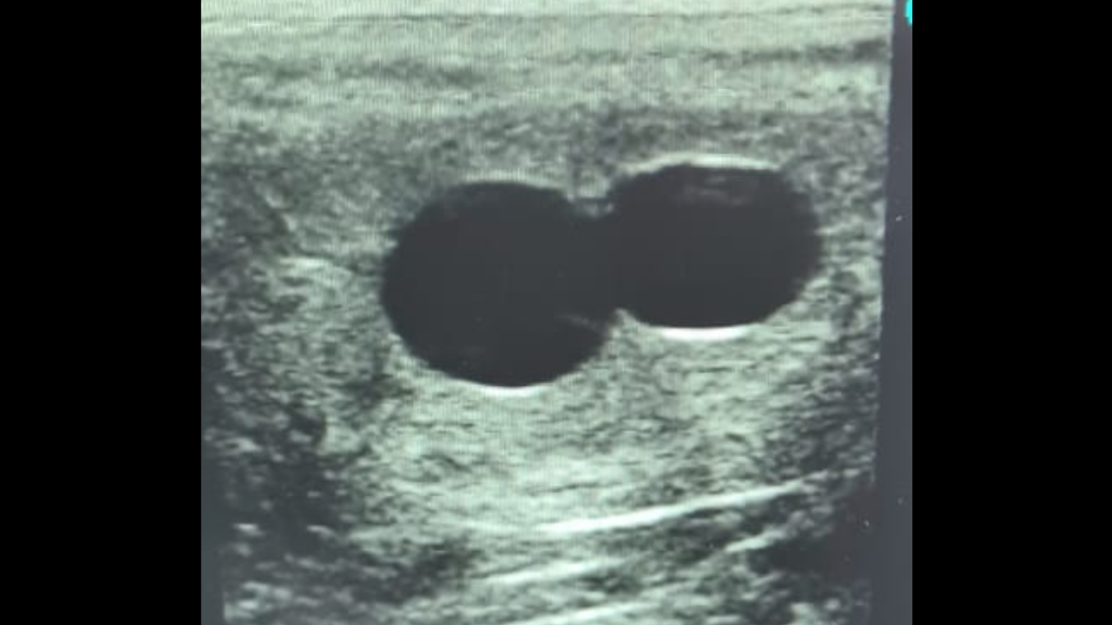
At approximately 60 to 70 days gestation, we employ transrectal ultrasonography to determine the gender of the developing foetus by identifying the location of a structure called the genital tubercle, which, in the developing foetus, is the structure which later becomes the clitoris in females and the prepuce (sheath) in males. The genital tubercle is identical in both male and female 60-70 day foetuses, however, the location of the structure is subtly different, which allows us to differentiate between the two. Beyond day 80 of pregnancy, the foetus is usually too far to ultrasound reliably via rectal ultrasound.
Transabdominal Gender Determination:
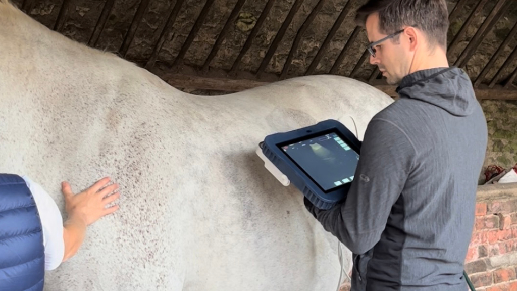
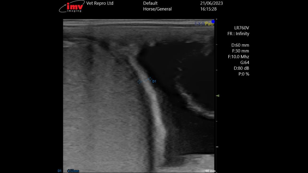
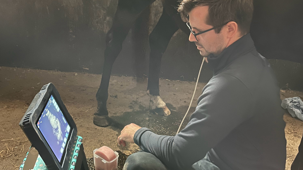
As the pregnancy progresses, from about between about 5 and 8 months, or sometimes sooner, we transition to transabdominal ultrasonography for foetal gender determination. This non-invasive technique allows for a more comprehensive visualisation of the developing foetus, providing breeders with more detailed information. The main structures we visualise to determine sex are the udder/prepuce (sheath), the perineum and the gonads themselves. The foetal udder in females and the foetal prepuce in males is located in the same location, between the hind legs and has a similar appearance on ultrasound scan. The key differentiating factor are the hyperechoic (bright white) teats seen associated with the udder in females. The perineum is also imaged in the same location for females and males; the female perineum is identified by a single line along midline under the tail head, while the male perineum is marked by two parallel lines, representing the urethra, which is the key differentiating factor here. The gonads are easily located in the foetal abdomen as they are particularly large during mid-gestation. While both early ovaries and testicles look very similar in size and shape, subtle differences can be seen in the tissue itself and colour-flow Doppler can be used to compare blood flow to the gonads as the pattern of blood flow differs between the two. Visualising several male or female foetal structures provides added certainty when determining foetal gender. It is also safer than the earlier approach as the technique is non-invasive (does not utilise rectal ultrasound) and is therefore Malton Equine’s preferred window. The procedure can be performed without clipping the hair off the abdomen in most cases and sedation is not required if the mare stands quietly.
Strategic Breeding Decisions:
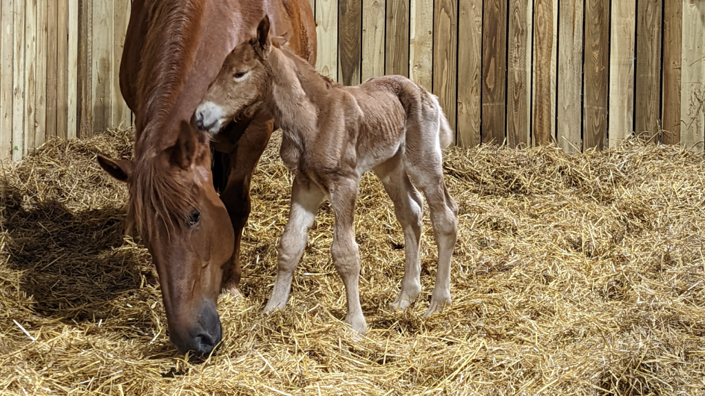
Accurate foetal gender determination plays a key role in strategic breeding decisions. It enables breeders to tailor their breeding programs, ensuring the desired genetic traits are perpetuated in subsequent generations. This information also allows for thoughtful management of bloodline diversity within a breeding operation.
Emergency veterinary attention for your horse may be required at any time of the day or night. We provide veterinary care 24 hours a day, 365 days a year to registered clients.

